🫀 الجهاز الدوري
🫀 The Circulatory System
💡 الفكرة الرئيسية: ينقل الجهاز الدوري الدم إلى جميع مناطق الجسم، وبذلك تحصل الخلايا على الأكسجين والمواد الغذائية اللازمة لحياتها، كما ينقل الفضلات الناتجة عن عمليات الأيض إلى أعضاء الإخراج للتخلص منها.
💡 Main Idea: The circulatory system carries blood to every region of the body, so cells receive the oxygen and nutrients they need to live, and it carries away the metabolic wastes produced by cells to the excretory organs for removal.
🔹 الربط بالحياة اليومية: يمكن تشبيه شبكة الأوعية الدموية بشبكة الطرق السريعة في مدينة: فكما تنقل الطرق السيارات المحمّلة بالبضائع إلى أحياء المدينة وتُخرج نفاياتها، تنقل الأوعية الدموية خلايا الدم المحمّلة بالأكسجين والمواد الغذائية إلى كل نسيج، وتُعيد حمل الفضلات وثاني أكسيد الكربون للتخلص منهما.
🔹 Connection to Daily Life: The network of blood vessels can be compared to a city's highway system: just as roads carry trucks loaded with goods to every neighborhood and carry waste back out, blood vessels carry blood cells loaded with oxygen and nutrients to every tissue, and carry wastes and carbon dioxide back out for disposal.
⚙️ وظائف الجهاز الدوري
⚙️ Functions of the Circulatory System
يجب أن تحصل خلايا الجسم على الأكسجين والمواد الغذائية باستمرار، كما يجب أن تتخلص من الفضلات الناتجة عن عمليات الأيض داخلها. ولأن معظم خلايا الجسم لا تتصل مباشرةً بالمحيط الخارجي، يقوم الجهاز الدوري بمهمة الوسيط: فهو يحمل الأكسجين من الرئتين والمواد الغذائية من الجهاز الهضمي إلى كل خلية، ويحمل ثاني أكسيد الكربون والفضلات الأخرى بعيدًا عنها. كما ينقل الهرمونات بين أعضاء الجسم، ويسهم في الدفاع عن الجسم ضد مسببات الأمراض عبر خلايا الدم البيضاء، وفي تخثر الدم لوقف النزيف عبر الصفائح الدموية، وفي تنظيم درجة حرارة الجسم.
Body cells must continuously receive oxygen and nutrients, and must get rid of the metabolic wastes they produce. Because most body cells have no direct contact with the outside environment, the circulatory system acts as the go-between: it carries oxygen from the lungs and nutrients from the digestive system to every cell, and carries carbon dioxide and other wastes away from them. It also transports hormones between organs, helps defend the body against pathogens via white blood cells, helps clot blood to stop bleeding via platelets, and helps regulate body temperature.
يتكون الجهاز الدوري من ثلاثة مكونات رئيسة: القلب (المضخة التي تدفع الدم)، الأوعية الدموية (الأنابيب التي يسري فيها الدم)، والدم نفسه (السائل الناقل).
The circulatory system has three main components: the heart (the pump that drives the blood), the blood vessels (the tubes the blood flows through), and the blood itself (the transport fluid).
📜 الشكل 1 — من الجثث إلى القلوب الصناعية: ساهمت دراسة الجهاز الدوري البشري تاريخيًا (بدءًا من تشريح الجثث) في تقدّم كبير في تقنيات الطب الحديث، وصولًا إلى تطوير القلوب الصناعية المستخدمة اليوم لمساعدة أو استبدال القلوب المصابة.
📜 Figure 1 — From Cadavers to Artificial Hearts: Studying the human circulatory system historically (starting with the dissection of cadavers) contributed to major advances in modern medical technology, leading up to today's artificial hearts, used to assist or replace damaged hearts.
📝 اختبر فهمك — الجهاز الدوري📝 Quiz — The Circulatory System
3 أسئلة3 Questions🩸 الأوعية الدموية
🩸 Blood Vessels
تضم الأوعية الدموية في جسم الإنسان ثلاثة أنواع رئيسة: الشرايين والشعيرات الدموية والأوردة، ولكل نوع منها بنية تلائم وظيفته.
The human body has three main types of blood vessels: arteries, capillaries, and veins, each built to match its own function.
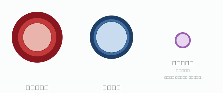
الشرايين: تنقل الشرايين الدم بعيدًا عن القلب. وتكون جدرانها سميكة ومرنة لتتحمل ضغط الدم الناتج عن انقباض القلب. تتكون جدران الشرايين من ثلاث طبقات: طبقة خارجية من نسيج ضام، وطبقة وسطى عضلية ومرنة، وطبقة داخلية ملساء (بطانة) تسمح بمرور الدم بسهولة.
Arteries: Arteries carry blood away from the heart. Their walls are thick and elastic to withstand the blood pressure produced by the heart's contractions. An artery wall has three layers: an outer connective-tissue layer, a middle muscular and elastic layer, and a smooth inner lining that lets blood flow through easily.
الشعيرات الدموية: تتفرع الشرايين إلى أوعية دموية أصغر فأصغر حتى تصل إلى الشعيرات الدموية، وهي أدقّ الأوعية الدموية وأكثرها عددًا. جدارها رقيق جدًا (بسماكة خلية واحدة)، ما يسمح بمرور الأكسجين والمواد الغذائية من الدم إلى الخلايا المجاورة، ومرور ثاني أكسيد الكربون والفضلات من الخلايا إلى الدم — وهذا هو موقع التبادل الفعلي بين الدم وخلايا الجسم.
Capillaries: Arteries branch into progressively smaller vessels until they reach capillaries, the smallest and most numerous blood vessels. Their wall is just one cell thick, which lets oxygen and nutrients pass from the blood to nearby cells, and lets carbon dioxide and wastes pass from the cells into the blood — this is where the actual exchange between blood and body cells happens.
1) قارن بين الأوعية الدموية الثلاثة الرئيسة (الشرايين والشعيرات الدموية والأوردة): ما الصفة المشتركة بينها؟
2) صف بنية جدران الشرايين.
1) Compare the three main blood vessel types (arteries, capillaries, and veins): what feature do they share?
2) Describe the structure of an artery's walls.
🔵 الأوردة
🔵 Veins
بعد أن يمرّ الدم عبر الشعيرات الدموية الدقيقة، يدخل إلى أوعية دموية أكبر تسمى الأوردة، وهي تحمل الدم من أعضاء الجسم عائدًا إلى القلب. يكون ضغط الدم في الأوردة منخفضًا نسبيًا (بعد أن فقد الدم كثيرًا من قوته الدافعة أثناء مروره عبر الشعيرات الكثيرة)، ولذلك تكون جدران الأوردة أرقّ من جدران الشرايين. وللمحافظة على تدفق الدم في اتجاه واحد نحو القلب رغم انخفاض الضغط، تحتوي الأوردة (خاصة في الأطراف) على صمامات تمنع رجوع الدم إلى الخلف.
After passing through the tiny capillaries, blood enters larger vessels called veins, which carry blood from the body's organs back to the heart. Blood pressure in veins is relatively low (the blood has lost much of its driving force passing through the many capillaries), so vein walls are thinner than artery walls. To keep blood flowing in one direction toward the heart despite the low pressure, veins (especially in the limbs) contain valves that prevent blood from flowing backward.
صِف أوجه الاختلاف بين الشرايين والأوردة.
Describe the differences between arteries and veins.
🤔 فكّر: الشكل 3 — ضع فرضية حول كيفية إسهام قطر الأوعية الدموية القريبة من سطح الجلد في تنظيم درجة حرارة الجسم.
🤔 Think About It: Figure 3 — Form a hypothesis about how the diameter of blood vessels near the skin's surface helps regulate body temperature.
📝 اختبر فهمك — الأوعية الدموية📝 Quiz — Blood Vessels
4 أسئلة4 Questions❤️ القلب
❤️ The Heart
القلب عضو عضلي، بحجم قبضة اليد تقريبًا، يقع في منتصف الصدر تقريبًا وينحرف قليلًا نحو الجهة اليسرى. يضخّ القلب الدم إلى جميع أجزاء الجسم، ويؤدي هذه المهمة باستمرار طوال حياة الإنسان وهو ينبض.
The heart is a muscular organ, roughly the size of a clenched fist, located near the center of the chest and shifted slightly to the left. It pumps blood to every part of the body, and keeps doing so throughout a person's life as it beats.
🔬 تركيب القلب
🔬 Heart Structure
يتكوّن القلب من أربع حجرات، وينقسم إلى جهتين (يمنى ويسرى). تُسمّى الحجرتان العلويتان الأذينين (الأذين الأيمن والأذين الأيسر)، وتُسمّى الحجرتان السفليتان البطينين (البطين الأيمن والبطين الأيسر). ويوجد بين كل أذين والبطين المقابل له صمام يفتح باتجاه واحد فقط (من الأذين إلى البطين)، ويمنع رجوع الدم إلى الخلف — بمبدأ مشابه لصمامات الأوردة.
The heart has four chambers and is divided into two sides (right and left). The two upper chambers are called the atria (right atrium and left atrium), and the two lower chambers are called the ventricles (right ventricle and left ventricle). Between each atrium and the ventricle below it is a valve that opens in one direction only (from atrium to ventricle) and prevents blood from flowing backward — the same principle as the valves in veins.
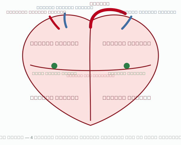
صف الاختلاف بين الصمامات الموجودة في القلب والصمامات الموجودة في الأوردة.
Describe the difference between the valves in the heart and the valves in veins.
🏃 هوية مرتبطة بعلم الأحياء: أخصائي فسيولوجيا التمارين
🏃 A Biology-Related Career: Exercise Physiologist
يدرس أخصائيو فسيولوجيا التمارين استجابات الجسم (ومنها الجهاز الدوري) للنشاط البدني، ويطوّرون برامج تمارين للرياضيين والأفراد لتحسين مستويات لياقتهم البدنية، ويراقبون هذه المستويات بمرور الوقت.
Exercise physiologists study the body's responses (including the circulatory system) to physical activity, design exercise programs for athletes and individuals to improve their fitness levels, and track those levels over time.
⚡ كيف ينبض القلب
⚡ How the Heart Beats
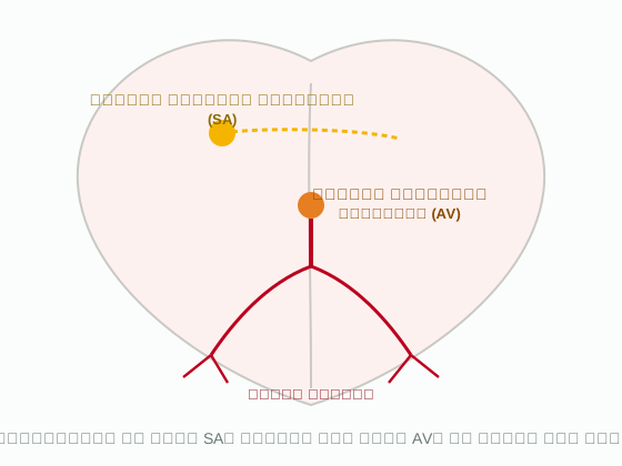
ينبض القلب على مرحلتين رئيستين: في المرحلة الأولى ينقبض الأذينان معًا، وفي المرحلة الثانية ينقبض البطينان معًا. يبدأ الانقباض من عقدة صغيرة تسمى العقدة الجيبية الأذينية (SA)، الواقعة في الجدار العلوي للأذين الأيمن، وتُرسل إشارة كهربائية منظّمة تنتشر عبر جدار الأذينين فينقبضان في آن واحد ويدفعان الدم إلى البطينين المرتخيين.
The heart beats in two main phases: in the first phase both atria contract together, and in the second phase both ventricles contract together. The contraction begins at a small node called the sinoatrial (SA) node, located in the upper wall of the right atrium, which sends an organized electrical signal that spreads through the atrial walls, so both atria contract at once and push blood into the relaxed ventricles.
عندما تصل الإشارة الكهربائية إلى العقدة الأذينية البطينية (AV) تتأخر جزءًا من الثانية، ليتسنى للأذينين إفراغ محتواهما من الدم بالكامل قبل أن تنقبض البطينات. ثم ينتشر السيال العصبي من العقدة الأذينية البطينية عبر ألياف موصلة تسمى ألياف بركنجي إلى عضلة البطينين، فينقبضان معًا بادئَين من قمة القلب.
When the electrical signal reaches the atrioventricular (AV) node, it is delayed for a fraction of a second, so the atria can empty their blood completely before the ventricles contract. The impulse then spreads from the AV node through conducting fibers called Purkinje fibers into the ventricular muscle, so both ventricles contract together, starting from the apex of the heart.
💓 النبضة
💓 The Pulse
يضخّ القلب نحو 70 مل من الدم في كل نبضة. وموجات ضغط الدم الناتجة عن ضربات القلب، والتي تنتشر عبر جدران الشرايين، تسمى النبض. يمكن قياس النبض بسهولة عند نقاط عديدة حول الجسم حيث تمر الشرايين قريبًا من سطح الجلد، كالمعصم والعنق، ويمكن عدّه بسهولة (نبضة واحدة لكل ضربة قلب).
The heart pumps about 70 mL of blood with every beat. The pressure waves produced by the heartbeats, which travel along the artery walls, are called the pulse. The pulse can be easily measured at several points around the body where arteries pass close to the skin's surface, such as the wrist and neck, and can be counted easily (one beat of the pulse for every heartbeat).
🩺 ضغط الدم
🩺 Blood Pressure
من أوضح المؤشرات على صحة القلب قياس ضغط الدم، الذي يُقاس عادةً على شريان الذراع. فإذا ارتفع ضغط الدم عن المعدل الطبيعي واستمر ذلك لفترة طويلة، فقد يشير ذلك إلى انسداد جزئي في الشرايين. وقد يشير الانخفاض الملحوظ في ضغط الدم إلى نزيف داخلي. تُقدَّر القراءة الطبيعية بـ 120 (الضغط الانقباضي) على 80 (الضغط الانبساطي).
One of the clearest indicators of heart health is blood pressure, usually measured on an artery in the arm. If blood pressure rises above the normal range and stays elevated for a long time, it may indicate partial blockage of the arteries. A noticeably low blood pressure reading may indicate internal bleeding. A normal reading is estimated at 120 (systolic pressure) over 80 (diastolic pressure).
📝 اختبر فهمك — القلب📝 Quiz — The Heart
4 أسئلة4 Questions🔁 تدفق الدم في الجسم
🔁 Blood Flow Through the Body
إذا تتبعت حركة الدم في الجسم، ستلاحظ أنه يدخل الأذين الأيمن، وينتقل إلى البطين الأيمن، ومن ثم إلى الرئتين، ويعود إلى القلب مرة أخرى ليدخل الأذين الأيسر، وينتقل إلى البطين الأيسر، ومن ثم يُضخّ إلى بقية الجسم عبر الشريان الأورطي. وبذلك ينقسم الجهاز الدوري إلى جهتين متكاملتين ومترابطتين: الدورة الدموية الرئوية (الصغرى) والدورة الدموية الجهازية (الكبرى).
If you trace the movement of blood through the body, you'll notice that it enters the right atrium, moves to the right ventricle, then to the lungs, returns to the heart to enter the left atrium, moves to the left ventricle, and is then pumped to the rest of the body through the aorta. The circulatory system is thus divided into two complementary, connected circuits: the pulmonary circuit and the systemic circuit.
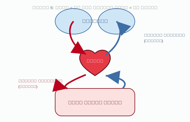
الدورة الدموية الرئوية (الصغرى): يبدأ الدم غير المؤكسج رحلته من الأذين الأيمن، وينتقل إلى البطين الأيمن، ويُضخّ منه إلى الرئتين عبر الشريان الرئوي. وعند وصوله إلى الشعيرات الدموية في الرئتين، يتخلص من ثاني أكسيد الكربون ويحمل الأكسجين، ثم يعود الدم المؤكسج إلى القلب عبر الأوردة الرئوية ليدخل الأذين الأيسر.
The pulmonary circuit (minor): deoxygenated blood begins its journey at the right atrium, moves to the right ventricle, and is pumped from there to the lungs through the pulmonary artery. On reaching the capillaries in the lungs, it releases carbon dioxide and picks up oxygen, then the oxygenated blood returns to the heart through the pulmonary veins to enter the left atrium.
الدورة الدموية الجهازية (الكبرى): بعد أن يمتلئ الأذين الأيسر بالدم المؤكسج، يمرّ إلى البطين الأيسر، ويُضخّ منه إلى بقية أجزاء الجسم عبر الشريان الأورطي. وأخيرًا، يعود الدم غير المؤكسج (المحمَّل بثاني أكسيد الكربون) إلى الأذين الأيمن عبر الوريدين الأجوفين العلوي والسفلي.
The systemic circuit (major): once the left atrium fills with oxygenated blood, it passes to the left ventricle, which pumps it to the rest of the body through the aorta. Finally, deoxygenated blood (carrying carbon dioxide) returns to the right atrium through the superior and inferior vena cavae.
📝 اختبر فهمك — تدفق الدم📝 Quiz — Blood Flow
3 أسئلة3 Questions🧪 استكشف الجهاز الدوري تفاعليًا (نموذج ثلاثي الأبعاد)
🧪 Explore the Circulatory System Interactively (3D Model)
جرّب النموذج التفاعلي أدناه: دوّره وكبّره لترى القلب النابض والرئتين والشرايين والأوردة والكليتين، وبدّل بين حالات الجسم المختلفة (راحة، طبيعي، رياضة، نوم) لترى كيف تتغير القياسات الحيوية التي درستها في هذا الدرس.
Try the interactive model below: rotate and zoom it to see the beating heart, lungs, arteries, veins, and kidneys, and switch between the body states (rest, normal, exercise, sleep) to see how the vital signs you studied in this lesson change.
↗️ افتح النموذج في صفحة كاملة ↗️ Open the model in a full page🩸 مكوّنات الدم
🩸 Components of Blood
💛 البلازما
💛 Plasma
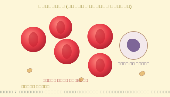
السائل الأصفر شبه الشفاف الذي ينقل مكونات الدم عبر مجرى الدم هو البلازما. تتكون البلازما من أكثر من 90% ماء، وحوالي 10% مواد أخرى ذائبة فيه، منها البروتينات والأملاح والفضلات والهرمونات والغازات الذائبة (كالأكسجين وثاني أكسيد الكربون).
The pale yellow, semi-transparent fluid that carries the components of blood through the bloodstream is plasma. Plasma is more than 90% water, with about 10% other substances dissolved in it, including proteins, salts, wastes, hormones, and dissolved gases (such as oxygen and carbon dioxide).
تقوم بروتينات البلازما بأنشطة حيوية مهمة: فبعضها (كالجلوبيولين) يساعد على مقاومة الأمراض والعدوى، وبعضها الآخر (كالفيبرينوجين) يساعد على تخثر الدم عبر تكوين الجلطات. كما يحافظ بروتين الألبومين على الضغط الأسموزي للدم، فيساعد في المحافظة على مستوى السوائل داخل الأوعية الدموية (توازن الماء في الجسم).
Plasma proteins carry out important vital activities: some (such as globulins) help resist disease and infection, while others (such as fibrinogen) help clot blood by forming clots. The protein albumin maintains the osmotic pressure of the blood, helping to maintain fluid levels inside blood vessels (the body's water balance).
اشرح وظائف البلازما.
Explain the functions of plasma.
🔴 خلايا الدم الحمراء
🔴 Red Blood Cells
خلايا الدم الحمراء أقراص مقعرة الوجهين، وتفقد نواتها أثناء نضجها. تحتوي كل خلية على نحو 250 مليون جزيء من الهيموغلوبين، الذي يرتبط به الأكسجين كيميائيًا (بواسطة الحديد الموجود فيه)، فتحمله خلايا الدم الحمراء إلى خلايا الجسم كلها.
Red blood cells are biconcave discs, and they lose their nucleus as they mature. Each cell contains about 250 million molecules of hemoglobin, which oxygen binds to chemically (via the iron it contains), so red blood cells carry it to every cell in the body.
⚪ خلايا الدم البيضاء
⚪ White Blood Cells
تكافح خلايا الدم البيضاء الأمراض، وتحمي الجسم من الميكروبات كالبكتيريا والفيروسات. عددها أقل بكثير من خلايا الدم الحمراء (نحو خلية بيضاء واحدة لكل 500-1000 خلية حمراء)، لكنها أكبر حجمًا منها، ويمكنها الخروج من الأوعية الدموية إلى الأنسجة المصابة لمهاجمة مسببات الأمراض مباشرة.
White blood cells fight disease and protect the body from microbes such as bacteria and viruses. They are far less numerous than red blood cells (roughly one white cell for every 500–1000 red cells), but larger in size, and can leave the blood vessels to enter infected tissues to attack pathogens directly.
🟡 الصفائح الدموية
🟡 Platelets
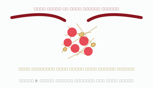
عند حدوث جرح في وعاء دموي، تتجمّع الصفائح الدموية عند موضع الجرح وتلتصق ببعضها، فيتحول بروتين الفيبرينوجين الذائب في البلازما إلى خيوط فيبرين غير ذائبة، تشكّل شبكة تحبس خلايا الدم الحمراء وتكوّن الجلطة الدموية التي توقف النزيف.
When a blood vessel is injured, platelets gather at the wound and stick together, causing the dissolved plasma protein fibrinogen to convert into insoluble fibrin threads, which form a mesh that traps red blood cells and forms the blood clot that stops the bleeding.
📝 اختبر فهمك — مكوّنات الدم📝 Quiz — Blood Components
4 أسئلة4 Questions🅰️ فصائل الدم
🅰️ Blood Types
وفق نظام فصيلة الدم ABO، توجد أربع فصائل رئيسة: A وB وAB وO، وتختلف فصيلة دم كل شخص بحسب مولدات الضد (antigens) الموجودة على سطح أغشية خلايا الدم الحمراء لديه. فإن كانت فصيلة الدم A، تحمل خلاياه مولد ضد A؛ وإن كانت B، تحمل مولد ضد B؛ وإن كانت AB، تحمل النوعين معًا؛ وإن كانت O، فلا تحمل خلاياه أي مولد ضد من النوعين.
Under the ABO blood type system, there are four main blood types: A, B, AB, and O, and each person's blood type depends on the antigens present on the surface of their red blood cell membranes. If the blood type is A, the cells carry the A antigen; if B, they carry the B antigen; if AB, they carry both; and if O, the cells carry neither antigen.
تُعدّ فصيلة الدم مهمة جدًا عند نقل الدم: يجب أن تتوافق فصيلة دم المتبرع مع فصيلة دم المستقبِل، وإلا تفاعلت الأجسام المضادة الموجودة في دم المستقبِل مع مولدات الضد الغريبة في الدم المنقول، فتلتصق خلايا الدم الحمراء ببعضها (تكتّل) وتسبب مشكلات خطيرة. الجدول 1 يبيّن مولدات الضد والأجسام المضادة لكل فصيلة، والفصائل التي يمكنها التبرع لها أو الاستقبال منها.
Blood type is very important in blood transfusions: the donor's blood type must be compatible with the recipient's, otherwise the antibodies in the recipient's blood react with the foreign antigens in the transfused blood, causing red blood cells to clump together (agglutination) and leading to serious problems. Table 1 shows the antigens and antibodies for each blood type, and which types can donate to or receive from each other.
| فصيلة الدمBlood Type | مولدات الضدAntigens | الأجسام المضادةAntibodies | يتبرع لـCan Donate To | يستقبل منCan Receive From |
|---|---|---|---|---|
| O | لا يوجدNone | A, B | O, A, B, AB | O |
| A | A | B | A, AB | A, O |
| B | B | A | B, AB | B, O |
| AB | A, B | لا يوجدNone | AB | O, A, B, AB |
الجدول 1: يُعدّ أصحاب فصيلة O مانحين لكل الفصائل (مانح عام)، وأصحاب فصيلة AB مستقبلين من كل الفصائل (مستقبل عام).
Table 1: people with type O are donors to all types (universal donor), and people with type AB can receive from all types (universal recipient).
➕➖ العامل الرايزيوسي Rh
➕➖ The Rh Factor
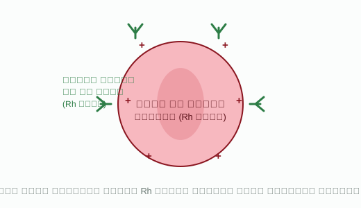
يُسمّى هذا العامل Rh لأنه اكتُشف أول مرة في دم قرد الريزوس. عندما يحمل الشخص مولد ضد Rh على سطح خلايا دمه الحمراء يُقال إن دمه موجب Rh، وعندما لا يحمله يُقال إن دمه سالب Rh. يمكن أن يحصل شخص سالب Rh على نقل دم موجب Rh مرة واحدة دون مشكلات خطيرة، لكن جسمه يبدأ عندها بإنتاج أجسام مضادة لعامل Rh، فإن تعرّض لدم موجب Rh مرة أخرى، تتفاعل هذه الأجسام المضادة مع الدم الجديد وتسبب تكتّل خلايا الدم الحمراء.
This factor is called Rh because it was first discovered in the blood of rhesus monkeys. When a person carries the Rh antigen on their red blood cells, their blood is said to be Rh-positive; when they don't, it is Rh-negative. An Rh-negative person can receive an Rh-positive transfusion once without serious problems, but their body then begins producing antibodies against the Rh factor, so if exposed to Rh-positive blood again, these antibodies react with the new blood and cause red blood cells to clump together.
يمكن أن يحدث هذا أيضًا في الحمل: إذا كانت الأم سالبة Rh والجنين موجب Rh (ورث عامل Rh من الأب)، فقد ينتج جسم الأم أجسامًا مضادة لعامل Rh إذا اختلط دم الجنين بدم الأم (كما قد يحدث أثناء الولادة). وفي حمل لاحق بجنين موجب Rh، يمكن لهذه الأجسام المضادة أن تعبر المشيمة وتدمّر خلايا الدم الحمراء للجنين، ما قد يسبب مشكلات صحية خطيرة. لذلك تُعطى الأم دواءً وقائيًا لمنع تكوّن هذه الأجسام المضادة.
This can also happen during pregnancy: if the mother is Rh-negative and the fetus is Rh-positive (inheriting the Rh factor from the father), the mother's body may produce Rh antibodies if the fetus's blood mixes with the mother's (as can happen during birth). In a later pregnancy with an Rh-positive fetus, these antibodies can cross the placenta and destroy the fetus's red blood cells, which can cause serious health problems. For this reason, the mother is given a preventive medication to stop these antibodies from forming.
📝 اختبر فهمك — فصائل الدم وعامل Rh📝 Quiz — Blood Types and the Rh Factor
4 أسئلة4 Questions⚠️ اضطرابات الجهاز الدوري
⚠️ Circulatory System Disorders
ترتبط كثير من اضطرابات الأوعية الدموية والقلب بأسلوب الحياة، وأهمها تصلّب الشرايين: تراكم رواسب دهنية دقيقة داخل جدار الشريان، ما يضيّق مجرى الدم ويقلّل مرونة جدار الشريان. يؤدي تراكم هذه الرواسب إلى ارتفاع ضغط الدم، وقد يؤدي انسداد شريان بالكامل إلى نوبة قلبية (توقف تدفق الدم عن جزء من عضلة القلب) أو سكتة دماغية (توقف تدفق الدم عن جزء من الدماغ)، ما قد يسبب تلف الأنسجة أو موتها بسبب نقص الأكسجين.
Many blood vessel and heart disorders are linked to lifestyle, the most important being atherosclerosis: the buildup of tiny fatty deposits inside an artery wall, which narrows the blood's pathway and reduces the artery wall's elasticity. The buildup of these deposits leads to high blood pressure, and a fully blocked artery can lead to a heart attack (blood flow stops to part of the heart muscle) or a stroke (blood flow stops to part of the brain), which can cause tissue damage or death from lack of oxygen.
💚 نصائح للوقاية: ممارسة الرياضة بانتظام، تناول غذاء صحي قليل الدهون، عدم التدخين، الحفاظ على وزن صحي، وقياس ضغط الدم بانتظام.
💚 Prevention tips: regular exercise, a healthy low-fat diet, not smoking, maintaining a healthy weight, and regularly checking blood pressure.
📝 اختبر فهمك — اضطرابات الجهاز الدوري📝 Quiz — Circulatory Disorders
3 أسئلة3 Questions📋 للمراجعة السريعة: افتح جداول ملخص الجهاز الدوري
📋 Quick review: Open the circulatory system summary tables
📝 امتحان الدورة الدموية📝 Circulatory System Exam
• عدد الأسئلة: 20 سؤالًا
• مدة الاختبار: 30 دقيقة
• لا يمكنك تغيير إجاباتك بعد التسليم، ولا إعادة الاختبار إلا بعد 24 ساعة من التسليم.
• تأكد من إجاباتك قبل التسليم! • Number of questions: 20 questions
• Exam duration: 30 minutes
• You cannot change your answers after submitting, or retake the exam until 24 hours after submission.
• Make sure of your answers before submitting!